Home
Welcome to Golden Lotus Herbs:
Introducing Ultra Premium Mono Floral Manuka Honey!
This Manuka honey has been tested twice in New Zealand and Germany by independent laboratories (Analytica and QSI Quality Services International) to ensure purity & potency.
We place great emphasis on delivering a quality that satisfies the highest of standards.

Golden Lotus Herbs is the home of the popular Lung & Throat Herbal Lozenges


Golden Lotus Herbs offers a vast selection of premium quality, natural combinations - Chinese Classics and herbal formulations of botanicals from around the world.
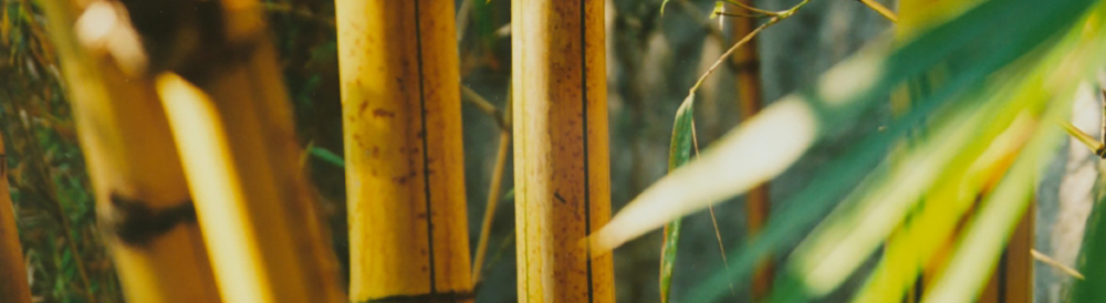
Here you can find a broad array of: traditional Chinese herbs, Western botanicals, and Ayurvedic herbs which are part of the diverse wealth of Traditional Herbal Heritage.
Our products are natural and organic or ecologically wild-crafted whenever possible. We supply concentrated liquid extracts, concentrated powdered extracts, classical herbal formulas, traditional, natural blends, fresh plant tinctures, glycerites, herbal lozenges, herbal creams, botanical oils, herbal throat sprays, and instant chai tonic teas.
For Shipping, Billing & Policies page click here
Golden Lotus Herbs is committed to preserving the rich heritage of herbal traditions and the promotion of wellness thru radiant health. We provide herbalists and health care practitioners with high quality selections of botanicals, custom formulations and natural herbal compounds, empowering practitioners to meet their client's particular needs.
Our product selection at Golden Lotus Herbs, includes: Western herbs, Chinese herbs, Ayurvedic and Amazon herbs, in single form or combinations of herbal blends. These products are available as fresh plant tinctures, liquid and concentrated powdered herbal extracts, glycerites, botanical oils, creams/ointments and topical sprays. Golden Lotus herbal apothecary also offers select nutritional supplements and innovative products from world class leaders in the professional wellness industry.
Product Updates!
PurBlack Shilajit: An Ayurvedic Treasure that must be experienced.
Golden Lotus Professional Consultation:
 At Golden Lotus Herbs, we work with healing arts practitioners, clinics, students and teaching institutions in maintaining their herbal dispensaries. Many practitioners who do not stock their own apothecaries have found our diverse collection and specialized services to be a valuable resource.
Our goal is to support your practice with impeccable service and quality botanicals. Our on-staff herbalists are available to answer questions and provide herbal research and reference material to qualified practitioners (Chinese Herbs, Western herbs, Nutritional Supplements, Etc.).
Don't hesitate to e-mail us - we are here to help. Golden Lotus Herbal Consultation E-mail: info@goldenlotusherbs.com
Golden Lotus Herb Quality Sourcing:
Chinese, Western, and Ayurvedic Herbs
 Our herbs are procured by skilled herbalists who carefully select only the best raw materials available. Organic and ethically harvested herbs are chosen whenever possible. Our premium herbal ingredients are manufactured by reputable manufacturers to the highest quality standards, yielding fresh, concentrated tea extracts with effective potency.
Golden Lotus supports eco-friendly agriculture and fair-trade practices which preserve local environments, herb farmers, and their ancient traditions of herb cultivation and processing.